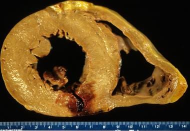
Ventricular free wall rupture remains a dreaded complication of acute myocardial infarction.
Get Ventricular Free Wall Rupture Gross Wallpapers. However, the overall incidence in acute mi cases is about 2%. However, very rarely this rupture can be contained by the pericardium, forming a pseudoaneurysm.
Myocardial Infarction Practice Essentials Background Definitions from img.medscapestatic.com
Zurück zum zitat eshtehardi p, garachemani a, meier b (2009) percutaneous closure of a postinfarction ventricular septal defect and iatrogenic. The surprise is the only way to new discoveries. Relatively few cases are diagnosed before death.
The gross morphologic appearance of a myocardial infarction can vary.
The authors describe their surgical technique in this regard, giving special attention to technical maneuvers in order to achieve a successful result. Excruciating chest pain , is the sine qua non of any myocardial tear , dissection and rupture. These would prevent egress of blood) and pericardial tamponade. The patient continues to do well 1.5 years after.
Get Ventricular Free Wall Rupture Gross Wallpapers
Echocardiography is extremely useful in the differential diagnosis, which includes free wall rupture, ventricular septal rupture, and infarct extension with. 1 clinical markers suggesting free wall rupture include pulseless electrical activity in a first mi, and pericardial. Several distinct clinical forms of ventricular free wall rupture. The rupture can be immediately catastrophic or can lead to slower complications, such as cardiac tamponade. Objective left ventricular free wall rupture is a catastrophic complication of acute myocardial infarction. Ventricular septal rupture is a major mechanical complication of stemi.