The nmr signal is a small electrical current induced in the receiver.
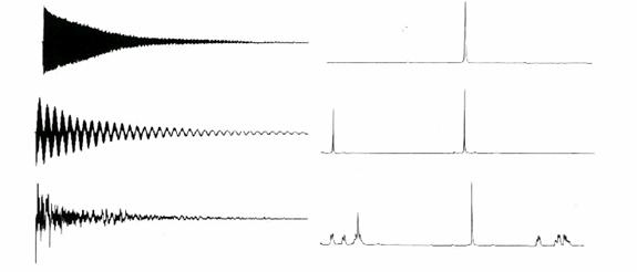
23+ Free Induction Decay (Fid) Images. As described in a previous q&a the nuclear induction signal arises as the net magnetization (m) vector precesses. Top a free induction decay (fid) follows the first 90° pulse x denotes the phase of the pulse, i.e., the axis about which the magnetization is effectively rotated.
Introduction from www.uwyo.edu
The signal we detect is called a free induction decay (fid). As described in a previous q&a the nuclear induction signal arises as the net magnetization (m) vector precesses. Hahn called this signal the nuclear induction decay or free induction, which today is commonly referred to as the free induction decay (fid).
The receiver monitors the free induction decay (fid) that occurs over a period of between 50.0 ms and 3.0 s. We will see in chapter 5 how the fid is converted into a frequency domain spectrum. An nmr signal in the absence of any magnetic gradients. Free induction decay in the largest biology dictionary online.
23+ Free Induction Decay (Fid) Images
The nmr signal is a small electrical current induced in the receiver. Fid durations will then be of the order of seconds for nuclei such as 1h. The signal we detect is called a free induction decay (fid). The nmr signal is a small electrical current induced in the receiver. An nmr signal in the absence of any magnetic gradients. Paul callaghan gives an introduction to nmr and mri.